On a morning, when we are still half asleep, and we put on moisturizer and enhance our eyelashes with mascara, we probably don’t spare much thought for the fact that these cosmetic products may well have been tested on animals. The safety of creams, makeup and lipsticks is often tested on animals so that we can look fresh and young without breaking out in a rash. Such testing has now been banned in Germany and Europe. But the effectiveness of medicines too is intensively tested on animals before they are introduced onto the market, different compounds and dosages are researched until a drug that is effective for humans with the least possible side effects has been created.
The team around Professor Dr. Heike Walles from the Institute of Chemistry at Otto von Guericke University Magdeburg is developing alternatives to these animal tests. With what is known as tissue engineering, targeted cells can be cultivated in the laboratory and thus human tissue produced outside of the body. Normally this is used to replace diseased tissue in patients, like, for example, cartilage. These days such endogenous cartilage implants are in certain cases even reimbursed by health insurance funds. However, in Magdeburg this tissue is not for use in transplants, but instead to enable risk assessments for cosmetics and medicines to be undertaken. DIN ISO-certified, with some of these tissue models, it is already possible to establish the biocompatibility - that is, the tissue compatibility - of certain substances and materials. Looking to the future with optimism, Professor Walles explains that “Our great aim is one day to be able to replace the majority of animal testing through tissue engineering and thus make research possible without animal suffering.” “To this end our aim is to develop many more models with which drugs, and in future medical products, can be tested for safety.”
Unfortunately the video is only available in German.
The focus of her research currently lies on applications for the airways. In the TIRAMISU research project, which is funded by the Federal Ministry of Education and Research to the tune of over 3.3 million euros, the biologists are cultivating pharyngeal tissue for endoscope manufacturers. This will enable them to develop devices that are able to recognize early changes in the mucous membrane. “Tumors often appear in the pharynx that are discovered far too late,” explains the scientist. “The earlier we detect them, the better they can be treated.” Often the trigger for the growth of tumors is a change in the microbiome, in other words, changes to the populations of bacteria that colonize our bodies. For this reason, the research team is developing different types of tissue in the laboratory: healthy tissue without a microbiome, healthy tissue with a healthy microbiome, healthy tissue with an altered microbiome, and tissue with an early-stage tumor. Using these tissue models, the aim is to train the endoscopes to detect the different signals using a laser. “At the end of the project we have to produce twelve tissue samples and give them to the manufacturer. The latter will then conduct the experiments without knowing which tissue is in front them,” explains Professor Walles. In the end, they have to achieve an accurate diagnosis for 90 per cent of the samples. If this double-blind test is successful, the chances are good that it will be possible to use the tissue models in future for other endoscopes and their approval.
To develop the models the scientists are using collagen - a protein from which our body constructs its connective tissue - and fibroblasts, which ensure that the collagen remains nice and firm. If it is already worn out or under too much strain, the fibroblasts break down the fibers and build new collagen. When we are young they do a better job, and by the end of our lives less so. This results in unwelcome wrinkles. In addition, they also pass important information on to other cells. “In the lungs, for example, the cells align themselves upwards towards the air. In our models, therefore, they are fed from below and have contact with air above. Thanks to the fibroblasts they know where up and down are,” explains Professor Walles. “We do the same with the skin. The cells feel where the skin comes from and then they begin to build themselves up like palisades - it is really fascinating.”
The models either consist of so-called cell lines, which, for example, are isolated from tumors. These are able to replicate themselves infinitely, but do not have all of the functions of the original cell. Or primary cells, that have been isolated from the tissue of patients, are used. However the research team is only able to work with these cells for a limited time. Skin cells, for example, divide ten times, then they die. If the skin models are built on these primary cells, then exactly the same thing happens. “It depends what we want to examine with the model. With the cell lines we can repeatedly conduct the same, standardized tests,” reports the biologist. “The tissue samples of the patients still have all of their functions but are not 100 per cent comparable with other samples. However, we are able to multiply them so that we can produce one hundred times the quantity of skin and can then freeze it at -150 degrees Celsius. We can conduct a lot of tests with that.
 Prof. Heike Walles (Photo: Jana Dünnhaupt / University of Magdeburg)
So far, what all of the models are lacking, among other things, are hairs and glands. Creating them would also be possible with the tissue engineering methods, but it is extremely time-consuming and expensive and not necessary for the current research at the University of Magdeburg. In summary, according to Professor Walles, their motivation is that “above all, we want to show that tissue engineering is suitable as an alternative to animal testing.” “With our predictions about the effectiveness of drugs, we are often even closer than is the case with animal testing. An already approved skin model for the testing of cosmetics does, for example, approximate the reactions that take place in the human body much more closely than testing on rabbits.” For the development of new tumor treatments, this is extremely important. Until now, mice had to be genetically modified or treated with certain substances to initiate tumor growth. Although the biological tissue of these tumors looks exactly the same, the background as to why the tumors have developed is different. “That is why they are often less informative,” points out Professor Walles. The researchers thus developed tissue models for the lungs for partners from the STIMULATE research campus. In future, newly developed electrodes should be able to systematically obliterate lung cancer, so that the tumors are killed off while causing the least possible damage to healthy tissue. These electrodes can be tested and optimized using these models without the need for animal testing.
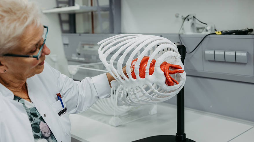
And it is not just medical products that can be improved with this technology. Surgeons can also work on their operative skills. The engineers are able, for example, to create a 3D model of a thorax using real patient data. This is printed out and the biological tissue is placed in the synthetic environment. A similar project is in planned in cooperation with the Faculty of Computer Science. Patient data will be used to simulate virtual reality aneurysms. This data could be used by Professor Walles to print out a skull with which procedures could be planned and practiced in advance specifically for a particular patient. “The first feedback from surgeons has definitely been positive. They cannot detect any difference to the real physiology; it feels like a real bone,” says Professor Walles. In the long term, the scientists also want to be able to print skin tissue. They would need special bio-inks, which they hope to develop in collaboration with colleagues from the Institute of Chemistry in the coming years. The big dream of Professor Walles, however, is to be able to work with fully synthetic materials. Alongside patients, the scientists rely on animal tissue to obtain collagen, for example from slaughterhouse waste. “The first synthetic models that we developed did not yet have the characteristics of biological materials. However, I am reasonably optimistic that we will come a large step closer to our objective in the next few years,” says Professor Walles.
 A printed heart is used to check whether the dimensions in the thorax are correct. Corresponding living tissue models are then constructed and integrated, thus creating a biophantom step by step as an alternative to the animal model. (Photo: Jana Dünnhaupt / University of Magdeburg)
To achieve this goal, the research team is benefiting from the expertise of other disciplines. Conversely, they also make their enormous expertise and the technological infrastructure for experiments available. To this end, in the Institute of Chemistry what are known as core facilities are being developed – a kind of co-working space for researchers, who are reliant on the expensive technology and expert knowledge for answers to their biological questions. Researchers from all over the world and from completely different disciplines can make use of these core facilities. “We ourselves have, of course, the greatest expertise in airway models. But that does not mean that we cannot work with a colleague on breast cancer,” says Professor Walles, who values the advantages of her interdisciplinary research field. “Ultimately we are a small cog helping to drive a bigger wheel. But together, even as a small university, we can make a lot of progress.”
Guericke facts
-
Since 2013 the sale of cosmetic products whose ingredients have been tested on animals has been banned.
-
The antibiotic Penicillin was first tested on rabbits in 1929, but showed no effect. Later tests on guinea pigs proved to be fatal.
-
In 2020, in German laboratories, tests were conducted on around 2.5 million animals. Genetic information was manipulated in over 900,000 animals. 19 per cent of animal tests were conducted to aid the manufacture or quality control of medical products.
