As the saying goes, “what I don’t know can’t hurt me”. “Right,” confirms Professor Dr. Daniel Behme, Chief Physician in Neuroradiology at Magdeburg's University Hospital. His professional interest lies in what are termed “incidental findings” that can derail an individual’s everyday life - for example, when an aneurysm is discovered in the brain. “Initially, most people do not notice a thing,” he says. It is only when, if for some reason or another, magnetic resonance imaging (MRI) or a computed tomography (CT) scan is carried out on the head, that by chance doctors discover an abnormal bulge in a blood vessel in the brain.
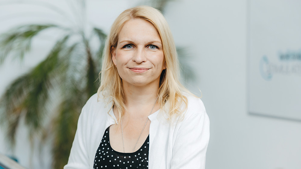
 In-vitro measurement of a flow diverter stent on the STIMULATE C-arm CT. Prof. Dr. med. Daniel Behme, Chief Physician of Neuroradiology at Magdeburg University Hospital, leads the clinical work package in the SOFINA project. (Photo: Jana Dünnhaupt / University of Magdeburg)
Strokes are a particular risk of an aneurysm. With every beat of the heart, blood is pumped through the blood vessels and thus into the bulge. If the vessel tears, it may trigger a life-threatening bleed or neurological symptoms such as paralysis, speech disturbances or reduced consciousness. As Professor of Interventional and Preventative Neuroradiology in the University Clinic for Radiology and Nuclear Medicine in Magdeburg, Daniel Behme has a wealth of knowledge in this field. The specialist in Neuroradiology moved here from the University of Göttingen in 2020. In his view, Magdeburg provides young academics with excellent career opportunities, for example with the STIMULATE research campus. This is a cooperation network between Otto von Guericke University Magdeburg, Siemens Healthineers AG, the STIMULATE Association and many partners from the medical and engineering sectors and industry.
STIMULATE acts as a magnet in Magdeburg’s Science Port. The research campus develops software and hardware for image-guided minimally-invasive therapies for treating cancer and vascular diseases - and also gives Professor Dr.-Ing. Sylvia Saalfeld just the conditions she needs for her research work. The scientist, who has worked at TU Ilmenau since April 2023, studied Computational Visualistics at Otto von Guericke University and wrote her post-doctoral thesis on the computer-aided treatment of intracranial aneurysms. At STIMULATE she had the opportunity to lead a research group working on medical image processing and visualization.
 Prof. Dr. Sylvia Saalfeld is responsible for the AI-based approaches in the research project. (Photo: Jana Dünnhaupt / University of Magdeburg)
Treatment of aneurysms
Which brings us back to the “incidental images” of brain aneurysms. “When they discover that they have a brain aneurysm, most people are extremely concerned,” says Daniel Behme. Many patients do not want to keep the aneurysm in their head, because they see it as a ticking time bomb. This analogy is also a valid one when it comes to the removal of the aneurysm. This is because the team of surgeons must approach the bulge in the blood vessel, which is surrounded by brain tissue, in the same careful way as disposal experts defuse a bomb. “With the traditional surgical method, the skull is opened up and the brain tissue moved aside in order to seal off the aneurysm using a clamp,” says the clinician, who goes on to explain that this process is increasingly being replaced by minimally-invasive interventions. But the more modern so-called endovascular interventions still require further optimization in order to provide safer care and treat patients in a more patient-friendly manner as well as contain the explosion in health care costs. The STIMULATE research campus, which was established in 2013 and lauded as one of Saxony-Anhalt’s cutting edge research venues, has now become an internationally respected pioneer.
Sylvia Saalfeld and her team are putting their skills to work in building the road to the future of medicine. This is because the analysis of the images produced by MRI and CT scans is playing an increasingly important role. “For example, we are looking at how 3D-models of the vascular defect can be extracted and simulated from this medical data, in order to be able to predict the most successful treatment method,” explains the computational visualistics expert. This is because the arrangement and composition of the vessels differ from patient to patient. “Furthermore, we know that aneurysms are themselves not the disease, but rather a symptom of a diseased blood vessel that is thin-walled in places and that would be better left untouched,” adds Daniel Behme, going on to explain a treatment method that targets this: “A so-called flow diverter - that is, a very finely woven, spiral-shaped stent - is placed in the blood vessel beneath the aneurysm. The blood flows past the aneurysm, with very little going into the bulge. The blood remaining in the bulge thromboses over time.”
 Original size of a flow diverter stent, which is used for minimally invasive endovascular treatment of intracranial aneurysms. (Photo: Jana Dünnhaupt / University of Magdeburg)
This means that this intervention is not completely without risk itself, as there is still a danger that the vessel wall will rupture. Daniel Behme demonstrates the guide wire, which is no thicker than a horsehair. It transports the stent on its tip. The wire is inserted into a vein in the thigh and pushed up into the brain. The “parent vessel”, as specialists call the vessel where the aneurysm has occurred, is generally a curved artery. Placing the stent in it with pinpoint accuracy is a delicate job for the neuroradiologist.
In future, the aim is for SOFINA to be able to support the treatment team and their research activities. The acronym stands for “Simulation-aided Optimization of Flow diverters for the treatment of INtracranial Aneurysms”. The Federal Ministry of Research is supporting this interdisciplinary project for a period of three years to the tune of 1.3 million euros. It is intended that the eventual result will be a new generation of flow diverters and a treatment planning software program that will optimize interventions. A further objective is to reduce the occlusion time; that is the time taken until the vessel is sealed by the formation of a thrombus.


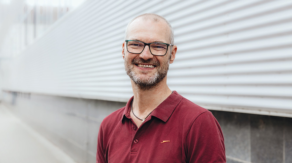
Photo left: Prof. Dr.-Ing. Gábor Janiga is responsible for the numerical blood flow simulation working group / Photo right: The virtual stent placement is realized by the working group of Prof. Dr.-Ing. (Photos: Hannah Theile / University of Magdeburg)
The project initiator is a member of the STIMULATE Association, Acandis GmbH. The Pforzheim-based company manufactures standardized neurovascular stents, which it hopes to be able to further improve with a view to offering more personalized medicine. The success of an intervention also depends, for example, on the material used to make the stent. For the next generation, the manufacturer hopes to test non-woven options, which will reduce the risk of complications.
The STIMULATE research groups, “Image Processing” led by Sylvia Saalfeld and “Medical Flows” led by Dr.-Ing. Philipp Berg, create 3D phantoms on the basis of medical data with which it is possible to simulate and predict the flow of blood outside of the human body. Interventions will stand a greater chance of success, according to Sylvia Saalfeld, if beforehand the patient’s exact circumstances can be simulated giving surgeons the opportunity for a rehearsal. During the intervention, the neuroradiologist can no longer influence a great deal. She shows us a phantom, a model of the affected vessel. In a block of plastic are an exactly recreated aneurysm and its parent vessel, in which the stent can be placed by way of a trial. The placing of the virtual stent and the flow of blood are simulated in the Institute of Mechanics under the leadership of Professor Dr.-Ing. Daniel Juhre and the Chair of Fluid Mechanics and Technical Flows at Otto von Guericke University under the leadership of Professor Dr.-Ing. Gabor Janiga.
 Silicone aneurysm models for risk-free testing of novel treatment approaches and validation of blood flow simulations. Left: 3D printed positive model with wall thickness, right: negative model in a silicone block (Photo: Jana Dünnhaupt / University of Magdeburg)
The two STIMULATE group leaders, Sylvia Saalfeld and Philipp Berg, emphasize that the collaboration with medical professionals is indispensable for the success of the project. They stress that the medical technology at their research campus has a high degree of practical relevance. Naturally, the Clinic for Neuroradiology at Magdeburg University Hospital is a member of the SOFINA alliance.
STIMULATE is the interface between industry and clinical practice. To ensure that the development of innovative medical products satisfies actual needs, the research campus has developed an Industry-in-Clinic platform and spun it off as a start-up with the support of the Federal Ministry of Research. “mediMESH - clinical insights” goes into clinics and creates an extensive database of medical workflows. It develops analytical tools to identify user needs and requirements as well as use-related risks. And it gathers digital feedback from medical experts on the newly developed products and prototypes.
“Our SOFINA project is one of the first major areas of activity for the new company,” says Philipp Berg, who heads the Young Talent Promotion and Careers support unit on the research campus. He welcomes mediMESH as another of the new companies making STIMULATE so attractive as a center for innovation and development.
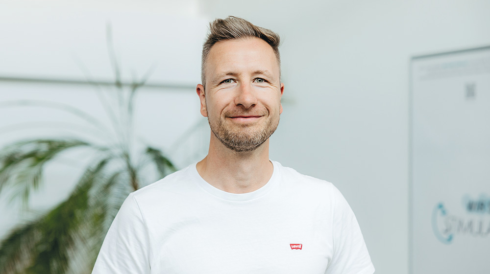
 PD Dr.-Ing. Philipp Berg heads the "Medical Flows" working group at the STIMULATE research campus and coordinates the research teams involved in the SOFINA project. (Photo: Jana Dünnhaupt / University of Magdeburg)
Guericke facts
- There is as much as a five percent chance of suffering from a cerebral aneurysm in an individual’s lifetime - thus one person in 20 is affected by this issue.
- Every year, the risk of rupture is around 2 percent. This rupture frequently leads to a life threatening stroke.
- An appropriate treatment has to be carefully evaluated. At present, the most common treatment method is a minimally-invasive intervention that leads, with the help of catheters, stents or coils, to the aneurysm being sealed off.
